EMR & ESD
2024-11-20 16:35:221. Technical details of EMR and ESD
How do EMR and ESD operate specifically, and what are their differences.
EMR - Endoscopic Mucosal Resection
EMR, or Endoscopic Mucosal Resection, is a technique that utilizes methods such as injection, suction, or ligation to separate flat protruding lesions (early gastrointestinal cancer, flat adenoma) and sessile polyps from their inherent layer, rendering them into pseudopolyps. These pseudopolyps are then removed using a snare or electrosurgery.
The indications for EMR include obtaining tissue specimens, gastrointestinal polyps, early-stage gastrointestinal cancer smaller than 2cm and confined to the mucosal layer, and tumors partially originating from the muscularis mucosae and submucosal layer.
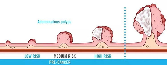
The specific operational methods of EMR in a broad sense include the following:
I. Polyp excision method, also known as submucosal injection-excision method, involves separating the submucosal layer, injecting liquid, and fully lifting the lesion through submucosal injection. If the lesion is less than 2cm, a snare can be used to encircle it, lift it along the wall, release the possibly clamped mucosal muscle layer, and then excise the lesion.

II. The transparent cap method (EMR with a cap, EMRC) involves installing transparent plastic caps of different specifications, planes, or inclinations on the end of the endoscope to attract and remove lesions. After submucosal injection of the lesion under endoscopy, the radiating snare is placed in the groove at the front end of the transparent cap, and the transparent cap is aligned with the lesion to be removed and inserted into the transparent cap. The snare is tightened to electrotome the lesion. Before electrotomy, the snare is also slightly loosened to allow the potentially affected muscularis propria to return to its original position.
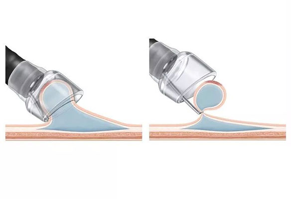
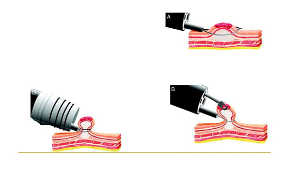
III. Endoscopic Mucosal Resection with Ligation (EMR with a Ligation, EMRL): Install the ligation device on the endoscope, align it with the lesion to be removed, and then use the rubber band to trap the lesion to form a sessile polyp. Subsequently, perform electrosurgery under the rubber band to remove the lesion, including the rubber band. This method is simple to operate, provides clear vision during the electrosurgery process, is easy to grasp the depth of resection, causes minimal local damage, has less surgical bleeding and complications, and is relatively safe.
ESD - Endoscopic Submucosal Dissection
ESD, or endoscopic submucosal dissection, is a new technology developed on the basis of EMR. Under endoscopy, high-frequency electrosurgical knives and specialized instruments are used to gradually dissect gastrointestinal lesions larger than 2cm (including early gastrointestinal tumors) from the normal submucosal layer beneath them, in order to achieve the goal of complete resection of the lesion.
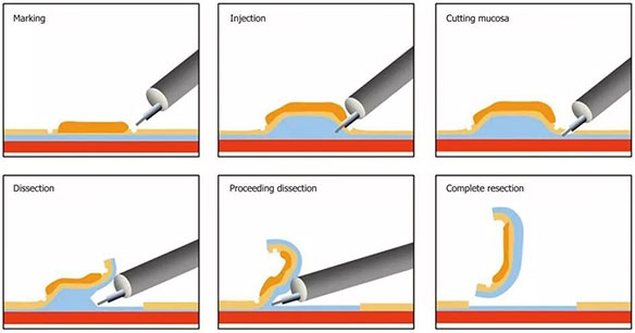
The specific operational steps are as follows:
I. Marking: Use a needle-shaped incision knife or argon plasma coagulation to electrically coagulate and mark the resection range at a distance of 0.5cm from the edge of the lesion;
II. Before submucosal injection, the clinically available liquids for submucosal injection include saline, glycerol fructose, sodium hyaluronate, etc;
III. Pre-incision of surrounding mucosa: Use a needle-shaped incision knife to incise the surrounding mucosa of the lesion along or outside the marked point, and then use an IT knife to incise the entire surrounding mucosa;
Ⅳ. Dissect the lesion. Depending on the location of the lesion and the surgeon's operational preferences, choose to use dissection instruments such as IT, Flex, or HOOK knife to dissect the lesion along the submucosal layer;
V. Wound management: Apply argon plasma coagulation to coagulate all visible small blood vessels on the wound surface to prevent postoperative bleeding. If necessary, apply hemostatic clips to clamp the blood vessels. 00:00 / 00:00
The indications for ESD include: gastrointestinal wide-based polyps and sessile polyps with a diameter greater than 2cm; early gastrointestinal cancer, which refers to cancer without lymphatic or hematogenous invasion or metastasis, and can be resected using ESD regardless of the location and size of the lesion; and gastrointestinal submucosal tumors with a diameter greater than 2cm.
2. Common consumables for EMR and ESD
Disposable high-frequency incision scalpel
The disposable high-frequency incision knife has good flexibility and conformability, and excellent suction performance. When used in conjunction with an endoscope and high-frequency electrotome, it can incise the surrounding tissues of digestive tract lesions.
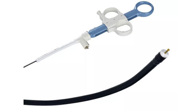
Disposable injection needle for endoscope
Disposable endoscopic injection needles can be used in conjunction with endoscopes to inject liquids such as physiological saline, glycerol fructose, etc. into the submucosa of the digestive tract, facilitating further separation of lesions.
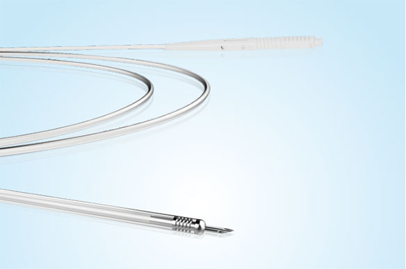
Disposable snare
A disposable snare is used in conjunction with an endoscope and a high-frequency generator, and is suitable for removing single or multiple polyps in the mucosal layer of the digestive tract. The size of the polyps can be captured by different caliber snare devices, with a diameter generally less than 2cm.
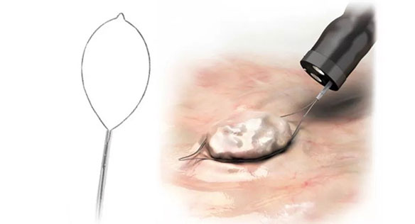
Transparent mucosal suction sleeve
Transparent mucosal suction sleeves are suitable for attracting and fixing small-sized lesions, separating them from normal mucosal tissue on the surface of the digestive tract, for further ligation of the lesions.
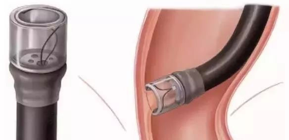
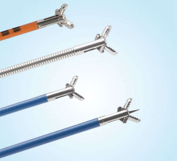
Disposable gripper
Disposable clamp, suitable for clamping and fixing lesions during snare ligation, to facilitate snare ligation and lesion resection.
3. Comparison and Summary of EMR and ESD
EMR and ESD share similar technical characteristics and are closely related. The disadvantage of EMR is that it is limited by the size of the lesion that can be resected under endoscopy (less than 2cm). If the lesion is larger than 2cm, segmental resection is required, and the incomplete treatment of the resection margin may lead to inaccurate postoperative pathology. However, ESD expands the indications for endoscopic resection, allowing for more complete resection of lesions larger than 2cm, making it an effective means for treating early-stage gastrointestinal cancer and precancerous lesions. Currently, both EMR and ESD are widely used in the resection treatment methods of digestive endoscopy.
EMR and ESD are the trump cards in endoscopic resection, having become crucial tools for minimally invasive treatment of early gastrointestinal cancer and precancerous lesions. I believe that in the future, EMR and ESD will create even greater medical value for public health.
Copyright © 2024 Changzhou Roga Medical Device Co.,Ltd. All Rights Reserved.
Powered by:ShuangXi

